Overview
The mPFC-HC memory system is comprised of regions of the medial temporal lobe, thalamus, and frontal cortex. Our lab conducts experiments to help further elucidate the basic anatomy, physiology, and neurobiological function of the mPFC-HC system using traditional anatomical tracing, viral tracing , confocal imaging, 3D reconstructions, in vivo electrophysiology, post-mortem high-resolution MRI, basic exploratory behavior, sophisticated cognitive tasks, and during sleep. Understanding the fundamental biology of the mPFC-HC system will provide the necessary foundations for making progress on understanding memory abilities, and when those systems go awry. These studies are funded, in part, by NIH grant R01 MH113626, and NIH grant R01 ES006189.
The mPFC-RE-HC system: Understanding higher order cortico-thalamo-cortical interactions in memory.
RE is a midline thalamic nucleus that projects to both the agranular mPFC, medial septum, and the stratum lacunosum molecular (slm) layers of CA1 and molecular layers of subiculum throughout the HC. A common mistake is to conceive of RE as a simple relay structure between the mPFC and HC, but a better conceptualization is as a higher order cortico-thalamo-coritcal system.
Our lab recently published a detailed synthesis on much of what is known about the RE at the anatomical and physiological levels in the context of memory that directly involves mPFC and HC functioning (Dolleman-van der Weel et al., 2019, LearnMem). Importantly, RE dysfunction has been established in several devastating disorders including Alzheimer’s disease, schizophrenia, and epilepsy. This is notable because the anatomy and physiology of RE has already provide key insights into the massive cognitive deficits seen severe Alzheimer’s, the development-related cognitive symptoms in schizophrenia, and the over-synchrony mechanisms epilepsy, but RE remains poorly understood.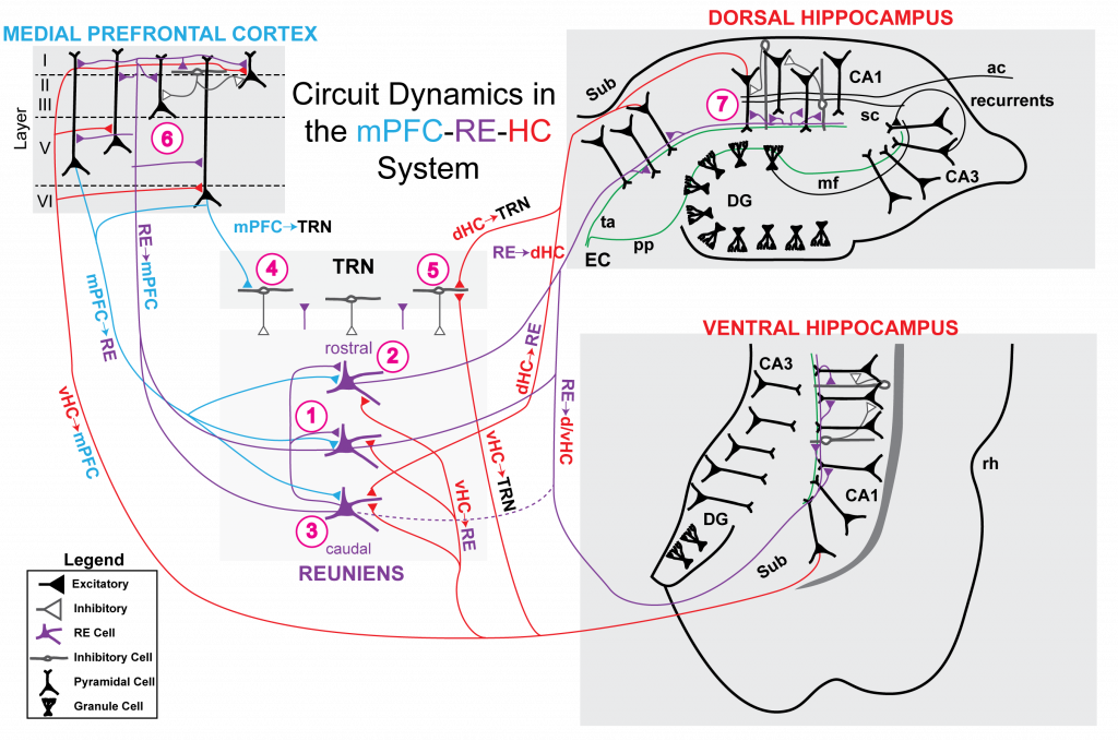
The mPFC-RE HC in humans
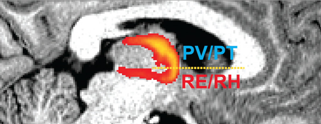
Little research has been conducted on the midline limbic thalamus in humans, a critical mediator of cognitive function. As in rodents, the anterior midline thalamus bi-directionally connects the mPFC and HC in primates. Based on connectivity, four regions of the anterior midline thalamus are thought to comprise the limbic thalamus including the RE, rhomboid nucleus (RH), peritoneal nucleus (PT), and paraventricular nucleus (PV). RE has been the most studied (in rodents) with roles in mnemonic tasks that depend on the mPFC, HC, and/or mPFC-HC interactions. However, little is known about the limbic thalamic nuclei in humans. This is due, in part, to the difficulty in identifying the limbic thalamus in imaging for the purpose of extracting structural and functional parameters. We addressed this problem using k-means clustering with probabilistic tractography in DWI data (Allen et al., 2016, SfN). This approach reveals two regions interconnected with the medial temporal lobe and agranular prefrontal cortex: (1) the ventral RE/RH region and (2) the dorsal PV/PT region. These anatomical findings accord well with evidence of these regions and their connectivity in the rodent brain.